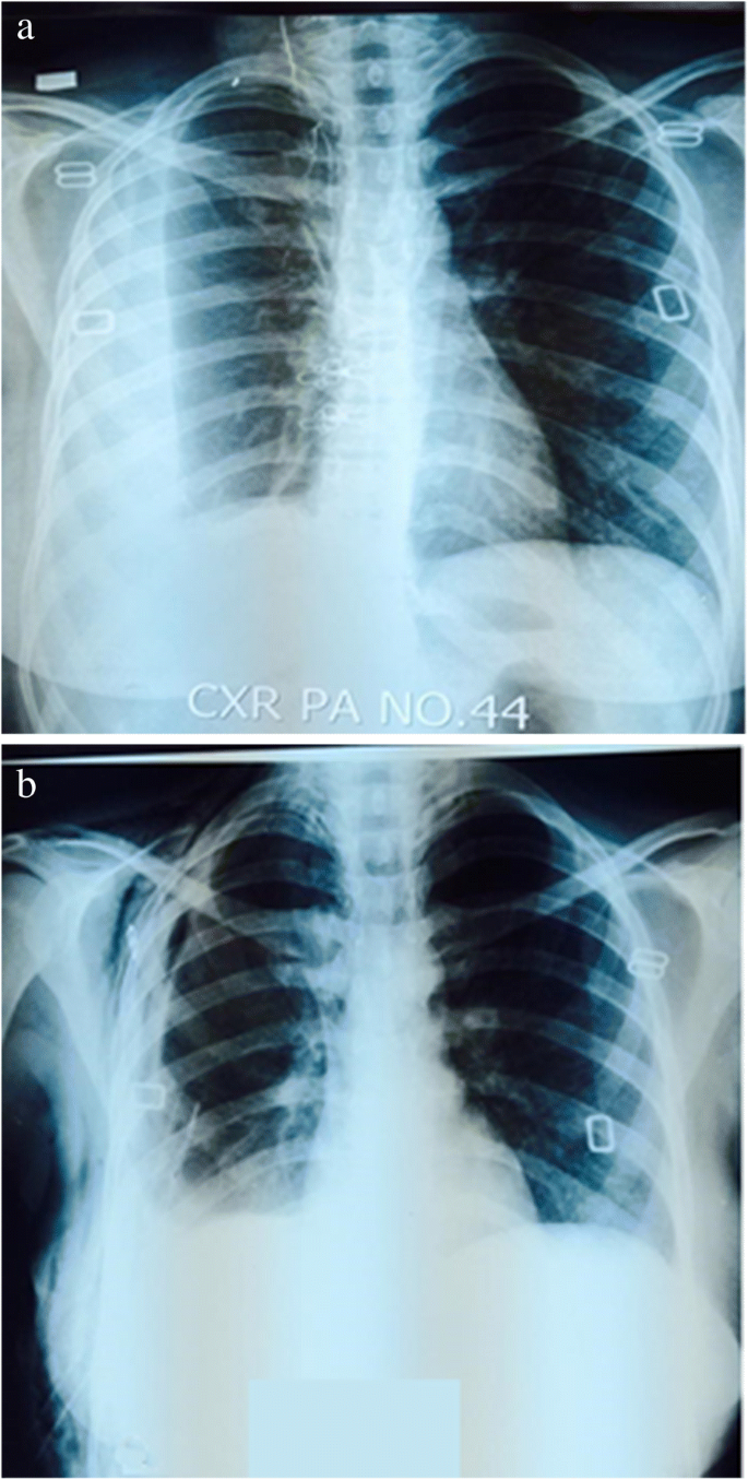
Loculated Pleural Effusion : Large Loculated Pleural Effusion 1 Of 3 / A loculated pleural effusion is the major radiographic hallmark of parapneumonic effusion or empyema (see fig.
Loculated Pleural Effusion : Large Loculated Pleural Effusion 1 Of 3 / A loculated pleural effusion is the major radiographic hallmark of parapneumonic effusion or empyema (see fig.. Most effusions start like this and can be easily missed. One of the most common reasons pleural effusion develops is due to. A pleural effusion is due to the manifestations of another illness.; What are the different appearances of pleural effusion? Pocus demonstrated a large right sided loculated pleural effusion with associated septations and surrounding consolidation suggestive of a parapneumonic effusion.
An ultrasound, chest computed tomograp. Loculated malignant effusions however, are inherently resistant to the usual approaches because of nonexpanding underlying lung. Pleural effusions are diagnosed in about 1.5 million individuals in the united states annually 1 . The pleural effusion is usually accompanied by parenchymal involvement; Other signs on the chest radiograph may suggest a malignant cause for the effusion.
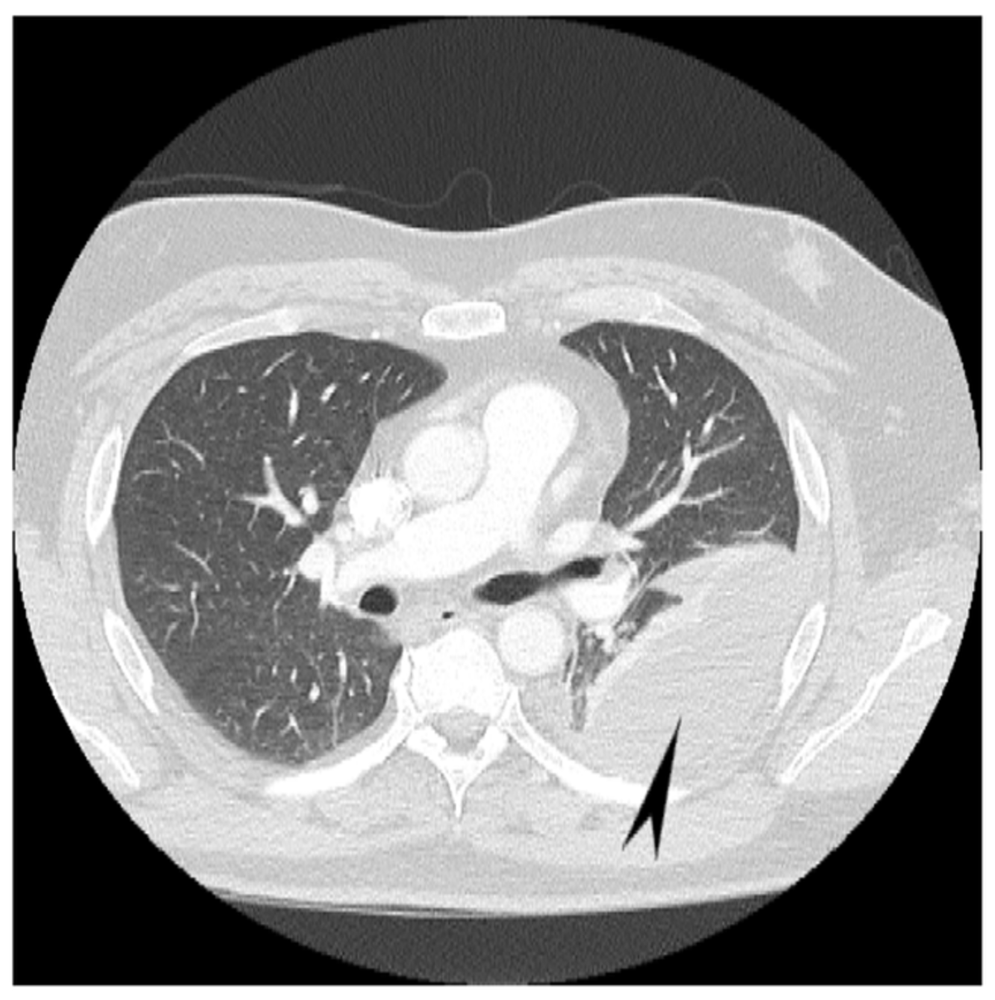
Cureus Hemorrhagic Pleural Effusion A Rare Presentation Of Vitamin K Deficiency In An Adult Patient from assets.cureus.com Pleural effusion that is confined to one or more fixed pockets in the pleural space. Loculated right pleural effusion with foci of atelectasis and consolidative changes concerning for pneumonia. Initial testing … lupus pleuritis and other causes of pleural effusions in lupus patients. Pleural effusions are currently addressed with aggressive antibiotic. In the context of a large effusion, mediastinal shift toward the side of the effusion should alert the clinician to the possibility of bronchial obstruction, which may. Pocus demonstrated a large right sided loculated pleural effusion with associated septations and surrounding consolidation suggestive of a parapneumonic effusion. Most effusions start like this and can be easily missed. Loculation most commonly occurs with exudative fluid, blood and pus.
One case of loculated pleural effusion secondary to amiodarone toxicity has been reported in literature.
Loculated malignant effusions however, are inherently resistant to the usual approaches because of nonexpanding underlying lung. Pleural effusions are currently addressed with aggressive antibiotic. Pleural effusions are diagnosed in about 1.5 million individuals in the united states annually 1 . Other signs on the chest radiograph may suggest a malignant cause for the effusion. Tell a friend about us, add a link to this page, or visit the webmaster's page for free fun content. If the fluid cannot be drained, the lungs aren't able to expand and oxygenate the blood sufficiently. A pleural effusion occurs when fluid fills this gap and separates the lungs from the chest wall. Loculation most commonly occurs with exudative fluid, blood and pus. Pleural effusion predominantly presents with breathlessness, but cough and pleuritic chest pain can be a feature. Surgical thoracostomy tube placement and radiologically guided catheter drainage are standard therapy for loculated pleural fluid collections. 1 article features images from this case 20 public playlist include this case Most effusions start like this and can be easily missed. Pleural effusions describe fluid between the two layer of tissue (pleura) that cover the lung and the lining of the chest wall.
Pleural effusion is when fluid fills this gap and separates the lungs from the chest wall. Cytological analysis of pleural fluid showed a negative result for malignant tumor cells. Normally, a small amount of fluid is present in the pleura. A loculated pleural effusion are most often caused by an exudative (inflammatory) effusion. The lack of specificity is mainly due to the limitations of the imaging modality.
Role Of Medical Thoracoscopy In The Management Of Multiloculated Empyema Bmc Pulmonary Medicine Full Text from media.springernature.com Fibrotic scar tissue may form in the pleural cavity (called loculation), preventing effective drainage of the fluid. Pocus demonstrated a large right sided loculated pleural effusion with associated septations and surrounding consolidation suggestive of a parapneumonic effusion. Treatment may fail if the catheter is not placed optimally within the loculation or if the fluid is hemorrhagic or fibrinous. Icu patients cannot sit up and the effusion layers posteriorly. Cytological analysis of pleural fluid showed a negative result for malignant tumor cells. Loculated effusions, defined as effusions that do not shift freely in the pleural space, occur when there are adhesions between the visceral and parietal pleura. Loculation most commonly occurs with exudative fluid, blood and pus. Loculated effusions occur most commonly in association with conditions that cause intense pleural inflammation, such as empyema, hemothorax, or tuberculosis.
A pleural effusion occurs when fluid fills this gap and separates the lungs from the chest wall.
Pocus demonstrated a large right sided loculated pleural effusion with associated septations and surrounding consolidation suggestive of a parapneumonic effusion. Causes of an exudative effusion are malignancy, infection, or inflammatory disorders such as rheumatoid arthritis. If the fluid cannot be drained, the lungs aren't able to expand and oxygenate the blood sufficiently. Loculated right pleural effusion with foci of atelectasis and consolidative changes concerning for pneumonia. The pleura are thin membranes that line the lungs and the inside of the chest cavity and act to lubricate and facilitate breathing. However, isolated pleural effusion has been reported. Other signs on the chest radiograph may suggest a malignant cause for the effusion. 681 views reviewed >2 years ago In chf effusions are bilateral and more on right. Enlarged mediastinal lymph nodes, possibly reactive. Most malignant effusions can be controlled by thoracentesis and/or closed thoracostomy tube drainage and sclerosis of the pleural cavity. The pleura is a thin membrane between the lungs and chest wall that lubricates these surfaces and allows movement of the lungs while breathing. A right loculated pleural effusion is still evident.
Pleural effusions are diagnosed in about 1.5 million individuals in the united states annually 1 . In general, pleural effusions can be divided into transudates (caused by fluid leaking from blood vessels) and exudates (where fluid leaks from inflammation of the pleura and lung). Surgical thoracostomy tube placement and radiologically guided catheter drainage are standard therapy for loculated pleural fluid collections. A pleural effusion occurs when fluid fills this gap and separates the lungs from the chest wall. Normally, a small amount of fluid is present in the pleura.
Pleural Effusion And Pneumothorax Springerlink from media.springernature.com Loculation most commonly occurs with exudative fluid, blood and pus. Pleural effusions are diagnosed in about 1.5 million individuals in the united states annually 1 . Loculated malignant effusions however, are inherently resistant to the usual approaches because of nonexpanding underlying lung. 681 views reviewed >2 years ago Most malignant effusions can be controlled by thoracentesis and/or closed thoracostomy tube drainage and sclerosis of the pleural cavity. In general, pleural effusions can be divided into transudates (caused by fluid leaking from blood vessels) and exudates (where fluid leaks from inflammation of the pleura and lung). 1 article features images from this case 20 public playlist include this case The pleura are thin membranes that line the lungs and the inside of the chest cavity and act to lubricate and facilitate breathing.
This can happen for many different reasons, including pneumonia or complications from heart, liver, or kidney disease.
Most effusions start like this and can be easily missed. If the fluid cannot be drained, the lungs aren't able to expand and oxygenate the blood sufficiently. Treatment may fail if the catheter is not placed optimally within the loculation or if the fluid is hemorrhagic or fibrinous. Fibrotic scar tissue may form in the pleural cavity (called loculation), preventing effective drainage of the fluid. Another reason could be as a side effect from cancer. The etiology of the pleural effusion determines other signs and symptoms. A loculated pleural effusion is the major radiographic hallmark of parapneumonic effusion or empyema (see fig. 681 views reviewed >2 years ago In the context of a large effusion, mediastinal shift toward the side of the effusion should alert the clinician to the possibility of bronchial obstruction, which may. Tell a friend about us, add a link to this page, or visit the webmaster's page for free fun content. Among the causes, pleural infection, heart failure, and malignancy are the most common. A pleural effusion occurs when fluid fills this gap and separates the lungs from the chest wall. This type of effusion is empyema unless proven otherwise.


0 Komentar